ECG or Electrocardiograph is an instrument for recording the changes of electrical potential occurring during the heartbeat used especially in diagnosing abnormalities of heart action.
More about ECG :
- Electrical voltages generated by the heart appear throughout the body.
- Sensitive instrumentation is used to measure the voltage at an electrode relative to some reference (e.g., another electrode).
- Measurements made at different locations will generate different recordings.
- Standard positions for electrode placement are needed so that clinical evaluations of ECG can be made.
ECG Lead Position
Lead I is the first of three standard limb leads (I, II, III). Limb leads measure cardiac depolarization in the frontal plane.
The negative electrode is connected to the RIGHT ARM. The positive electrode is connected to the LEFT ARM.
When an action potential starts on the right and proceeds toward the left side of the heart, a positive inflection will be seen in Lead I.
The negative electrode is connected to the RIGHT ARM. The positive electrode is connected to the LEFT ARM.
When an action potential starts on the right and proceeds toward the left side of the heart, a positive inflection will be seen in Lead I.

Lead II is one of the most common leads used for ECG monitoring. Normal rhythms present with a prominent P wave and a tall QRS.
The negative electrode is connected to the RIGHT ARM. The positive electrode is connected to the LEFT LEG.
The negative electrode is connected to the RIGHT ARM. The positive electrode is connected to the LEFT LEG.

Lead III is the last of the three standard limb leads.
The negative electrode is connected to the LEFT ARM. The positive electrode is connected to the LEFT LEG.
In all the limb leads, the electrodes may be positioned close to the torso. For convenience, they are often placed at the shoulders and hips.
As the electrodes are moved onto the torso, changes will occur in the measured ECG. This is one of the common sources of “differences” in the appearance of ECG's taken by our cart products compared to those taken by bedside monitoring products.

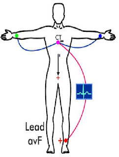
Lead aVF is the last of the three additional frontal plane leads.
The negative electrode is the central terminal. The positive electrode is connected to the LEFT LEG.
Notice that this vector bisects the angle formed by leads II and III.
The negative electrode is the central terminal. The positive electrode is connected to the LEFT LEG.
Notice that this vector bisects the angle formed by leads II and III.

Lead aVL is another of these three additional frontal plane leads.
The negative electrode is the central terminal. The positive electrode is connected to the LEFT ARM.
This one is slightly more difficult to visualize, but this vector bisects the angle formed by leads I and III … when we envision lead III being oppositely deflected.
The negative electrode is the central terminal. The positive electrode is connected to the LEFT ARM.
This one is slightly more difficult to visualize, but this vector bisects the angle formed by leads I and III … when we envision lead III being oppositely deflected.
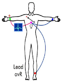
 Lead aVR is the first of these three additional frontal plane leads.
The negative electrode is the central terminal.
The positive electrode is connected to the RIGHT ARM.
The “a” denotes an augmented potential, accomplished by disconnecting the central terminal attachment from the explored limb.
We will see later that these additional frontal plane leads are routinely calculated from the standard limb leads (I, II, III). One of our primary purposes here is to notice that this vector (although oppositely deflected) bisects the angle formed by leads I and II.
Other Leads
An additional set of six electrodes, labeled V1-V6 or C1-C6, are placed on the chest and used to form what are known as the precordial leads.
The positive electrode is connected to the chest. The negative electrode is the central terminal.
The precordial leads measure the ECG potential in the transverse, or horizontal, plane.
The positive electrode is connected to the chest. The negative electrode is the central terminal.
The precordial leads measure the ECG potential in the transverse, or horizontal, plane.
Reference :
www-medlib.med.utah.edu/kw/ecg
www.ecglibrary.com
www.anaesthetist.com/icu/organs/heart/ecg
www.heartcenteronline.com


1 komentar :
Ijin share di blog saya, dgn sumber lengkap dicantumkan
Post a Comment